Each shade of colorrepresents different levels of attenuation. To the right of the image is agrayscale bar with numbers at the top, middle, and bottom.

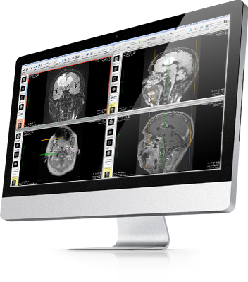
The image screen provides an abundance of information about the imagecurrently being viewed. This allows the user to change between series by left clicking the desiredseries, or open a new window (by right clicking the image) in tandem with the currentseries. There is now a bar with images that runs down the left sideof the screen. Once images are imported, the patient and image series can be selected andviewed in the 2D viewer mode.2D Viewer Mode:3įigure 4.In 2D viewer mode, it is noticeable that the Apple toolbar stays the same, whilethe OsiriX Toolbar changes. Clicking on themwill grant access to most of the functions that OsiriX has to offer.1įigures 2-3.There are two methods to adding images to the database, one is to click on ‘Import’ in theOsiriX Toolbar and select the file/images ready for importing, or click on ‘File’ from theApple Toolbar then click ‘Import’ ‘Import from file’ and select the file/files ready forimporting. They are: OsiriX, File, Edit,Format, 2D Viewer, 3D Viewer, ROI, Plugins, Window, and Help. Since there are no images in the Local Database yet, nothing is showingup in the lower windows.In the Apple Toolbar there are objects to click on. Underneath the OsiriX Toolbar is the Local Database.This contains a list of all the patients/specimens that have been imported into OsiriX.The bottom left of the screen displays all of the series of images for the selected patient.The bottom right of the screen displays the first image of the selected series of theselected patient. The OsiriX Toolbar is located beneath the Apple Toolbar, andruns the length of the window. The Apple Toolbar runs across the top ofthe screen all the time. This guide is aimed at explaining some of the basic, and intermediate uses ofOsiriX.Starting off:The first time the OsiriX application is run, there will be nothing in the Database.The window should look something like this:Figure 1.The screen is visibly divided into five sections. It can even save images in another format,making it easy to use desired or key images in presentations, publications, and forresearch. OsiriXis not limited to medical viewer formats such as CT or MR, it can also view images savedas jpg, tiff, mov, NIfTI, and much more.
#Osirix windows how to#
It is an easy to use application, and easy to learn how to use as well.
#Osirix windows software#
Introduction:OsiriX is an open source DICOM viewing software developed for Maccomputers. Jacqueline F WebbDepartment of Biological ScienceUniversity of Rhode IslandTimothy July 2010Funded by NSF Grant # 0843307 to J.F. A Guide to OsiriXBy Timothy AlbergLab of Dr.


 0 kommentar(er)
